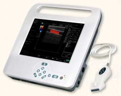
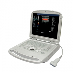
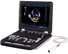
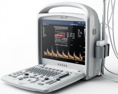
Features:
| 1. Streamline design, 4.5 kgs net weight |
| 2. Boot-up within 30 seconds |
| 3. 2+ hours battery working time |
| 4. 12.1 inch high resolution LED monitor |
| 5. Specific PICC puncture and placement software |
| 6. PICC specialty probe |
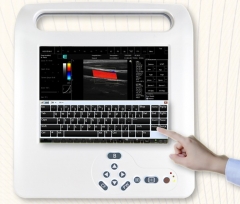
| 7. Touch-screen operation |
| 8. Touch and pop-up electronic keyboard |
| 9. 12 languages |
| 10. 18 user-interface skins |
Imaging Modes
B, B|B, 4B, B|M, M
Color Doppler (CFM)
Power Doppler (PDI)
Directional Power Doppler (DPDI)
Pulsed Wave Doppler (PWD)
B+PWD (Duplex)
B+CFM/PDI/DPDI+PWD (Triplex)
High Pulse Repetition Frequency (HPRF)
Tissue Harmonic Imaging (THI)
Continuous Doppler (CW)
Scanning Method
Electronic linear, electronic convex, electronic micro-convex, scanning depth: 2-24cm
Doppler ultrasound scanner
- PRF variable: 0.5-9 kHz
- Wall filter settings: 3 steps (5%, %10%, 15% PRF)
- Angle steering for linear transducers: ±10°
- Real-time spatial filter: 4 values
- CFM palette>10 maps
- PDI palette>10 maps
- B/Color priority control
- Color threshold control
- CFM baseline control
- Doppler frequency selection
- CW
- Color frame averaging
- Transparent Color Mapping (TCM)
Pulsed Wave Doppler
- PRF variable: 1-10 kHz
- Wall filter settings: 16 steps (2.5%-20% PRF)
- Angle steering for linear transducers: ±10°
- Real-time trace line with automatic calculation of spectrum parameters
- Stereo sound: volume control
- PWD palette>10 maps
- Doppler frequency selection
Processing
- High Line Density scan mode for better resolution
- 8 sliders TGC Control
- Dynamic range>120 dB
- Overall gain control
- M - mode sweep speed control
- Acoustic power control
- Variable frame averaging
- Brightness, contrast
- Advanced gamma control
- Scan direction, rotation, up-down controls
- Negative / positive control
- Echo enhancement control
- Noise rejection function
- Speckle reduction
Image and video
AVI, JPG, BMP, PNG, TIF, DCM (DICOM)
General Measurements and Calculations
Distance, Length, Area, Circumference, Volume, Angle, Stenosis %, A/B Ratio, Velocity, Pressure Gradient (PG), Acceleration, Resistivity Index (RI), Heart Rate, Velocity Time Integral (VTI), etc.
Measurements and Calculations Software Packages
Obstetrics, Gynecology, Abdominal, Urology, Endocavity, Vascular, Cardiology, etc.
Optional 3D /4D module
Expansion interfaces
VGA, TV Interface
USB2.0 Interface
RJ-45 Network interface
Support DeskJet printer, LaserJet printer, video printer
Imaging Technology:
Continuous High-precision Digital Beam-former
Dynamic Frequency Integration Imaging
High-precision Dynamic Receiving Focus
Super Wide-band Imaging Technology
Self-adaptive Image Optimization Processing
Multi-beam Imaging
Automatic Flow Volume Analysis
Compound Imaging
Panoramic Imaging
Self-adaptive Vascular Imaging
Self-adaptive Doppler Imaging
THI (Tissue Harmonic Imaging)
TDI (Tissue Doppler Imaging)
Image Processing
Pre-processing: 8-segment TGC
gross gain
dynamic range
gray map
smooth
acoustic power adjustment
scanning angle selection
Post-processing: edge enhancement
frame correlation
line correlation
γ-correction
contrast
brightnes
Accessories:
3.5 MHz convex probe
5.0 MHz micro-convex probe
6.5 MHz transvaginal probe
7.5 MHz linear probe
Biopsy guide
Luxury mobile trolley
Video/laser printer